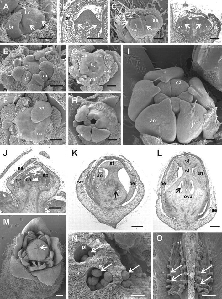
Fig. 4.

Floral development in Cedrela fissilis. In (E–I) the sepal, petal and/or part of the stamen primordia were removed for a better visualization of the inner structures. (A) Initiation of the abaxial sepal primordia (arrows) in a stage 3 meristem of a lateral male flower of a cymule in lateral view. (B) Longitudinal microtome section of a stage 3 floral meristem, where the abaxial (left arrow) and adaxial (right arrow) sepal primordia are differentiating. (C) Formation of petal primordia (arrows) in the second whorl, intercalary to the first whorl sepal primordia in stage 5. (D) Longitudinal section of a stage 5 flower meristem; the right and left arrows point to abaxial and adaxial petal primordia, respectively. (E) Formation of stamen primordia alternating with the petal primordia at stage 6. (F) Onset of carpel primordia alternating with the stamen primordia at early stage 7. (G) Stage 7, showing the initiation of the placenta as an invagination in the base of each carpel primordium (arrow). (H) Late stage 8, with carpel primordia elongating and undergoing post-genital fusion between carpels to form the ovary. (I) Late stage 10. As the carpels elongate, they displace the developing anthers. At the end of stage 10, the style and stigma begin to differentiate. (J) Longitudinal section of a terminal floral meristem at stage 7. (K) Longitudinal section of a stage 10 meristem of a functionally female flower. The arrow points to the developing ovule primordia. (L) Longitudinal section of a stage 11 male flower, with degenerating ovule primordia (arrow). (M) Stage 12, with a male flower at anthesis. The scars resulting from the post-genital fusion of the carpels remains visible (arrow). (N) Stage 11, female flower. Ovary locules with the top removed to expose developing ovule primordia (arrows). (O) Stage 12, male flower. Longitudinal view of the two exposed ovary locules (dorsal wall removed) showing degenerated ovules (arrows). an, anther; br, bracts; ca, carpel; fm, floral meristem; ova, ovary; pe, petal primordium; pl, placenta; se, sepal primordium; sp, stamen primordium; st, stigma; sl, style. Scale bars: A–D = 250 µm; E–G, M = 500 µm; K = 600 µm; L = 800 µm; N = 200 µm; O = 400 µm.
