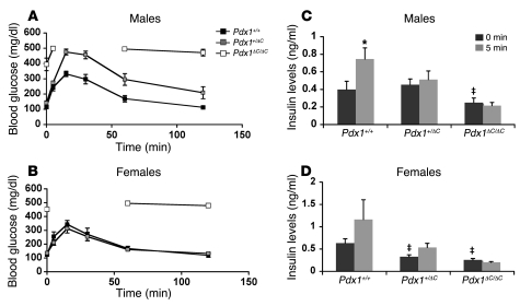
Figure 3. Impaired glucose tolerance and insulin secretion in Pdx1ΔC mice.
(A and B) Glucose tolerance in male (A) and female (B) mice. Intraperitoneal glucose tolerance tests using 2 g glucose/kg body weight performed on 3- to 4-week-old mice (n = 4–7 per group; ANOVA; male Pdx1+/+ versus Pdx1+/ΔC, P = 0.0037; male Pdx1+/+ versus Pdx1ΔC/ΔC, P < 0.0001; male Pdx1+/ΔC versus Pdx1ΔC/ΔC, P < 0.0001; female Pdx1+/+ versus Pdx1+/ΔC, P = NS; female Pdx1+/+ versus Pdx1ΔC/ΔC, P <0.0001; female Pdx1+/ΔC versus Pdx1ΔC/ΔC, P < 0.0001). Glucose levels in Pdx1ΔC/ΔC mice rose above 500 mg/dl, the limit of detection of the handheld glucometer. (C and D) Blunted acute insulin release in Pdx1ΔC/ΔC animals. Insulin levels were measured from whole blood collected at 0 and 5 minutes after intraperitoneal glucose injection (2 g glucose/kg body weight). Both male (C) and female (D) mice were 3–4 weeks of age (n = 4–7 animals per group). *P < 0.05 compared with level at 0 minutes after injection of same genotype; ‡P < 0.05 compared with level at 0 minutes after injection of wild-type mice.

