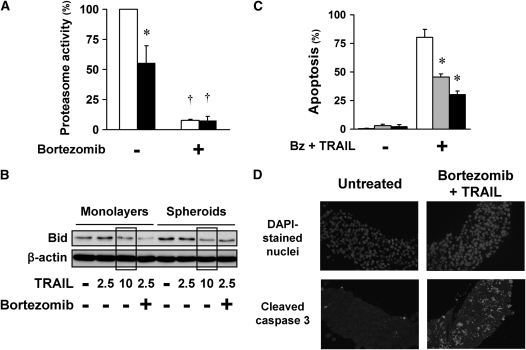
Figure 2.
Apoptotic resistance of spheroids to bortezomib plus TRAIL is not due to the limited diffusion of reagents into the spheroids. (A) Bortezomib inhibits the proteasome activity equally in A549 monolayers (open bars) and spheroids (solid bars). Under baseline conditions, proteasome activity was lower in A549 cells in spheroids than in monolayers. After exposure to bortezomib (100 nM) for 4 hours, proteasome activity in both monolayer and spheroid was equally inhibited (*different from monolayer; †different from baseline; mean ± SD; n = 3; P < 0.01). (B) TRAIL induced similar Bid cleavage in monolayers and spheroids. A549 monolayers and spheroids were exposed to TRAIL (2.5, 5, or 10 ng/ml) with or without bortezomib (100 nM) for 16 hours. TRAIL-induced Bid cleavage was measured by immunoblot as a decrease in full-length Bid. TRAIL 2.5 and 10 ng/ml induced similar Bid cleavage in A549 monolayers and spheroids (see boxed areas). After combination therapy, Bid cleavage is enhanced in monolayers compared with spheroids, consistent with an enhanced Bid cleavage seen after apoptosis and widespread activation of caspases (2). (C) Microspheroids demonstrate apoptotic resistance similar to that of spheroids. A549 cells grown as monolayers (open bars), microspheroids (shaded bars), and spheroids (solid bars) were exposed to 100 nM bortezomib (Bz) plus 1 ng/ml TRAIL for 24 hours, and then studied for apoptosis after Hoechst 33342 nuclear staining. Despite their difference in size and shape, and the shorter diffusion distance in the microspheroids, microspheroids and spheroids showed similar apoptotic resistance to bortezomib and TRAIL (*different from monolayer; mean ± SD; n = 3; P < 0.01). (D) Apoptotic cells are distributed evenly throughout the spheroids. A549 multicellular spheroids were treated with bortezomib (100 nM) plus TRAIL (1 ng/ml) for 24 hours, and then prepared for immunohistochemistry. Nuclei of all cells are indicated by DAPI staining; apoptotic cells are those with cleaved caspase 3. After exposure to bortezomib plus TRAIL, apoptotic cells are located throughout the spheroids, not in any one particular region (negative control with no primary antibody showed no staining; positive control, thymus, showed characteristic staining for apoptotic cells).

