Figure 2.
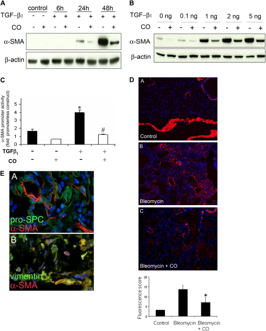
CO suppresses α–smooth muscle actin (α-SMA) expression in vitro and in vivo. Fibroblasts treated with TGF-β1 (5 ng/ml) were subsequently exposed to CO (250 ppm) or control incubator air. (A) Cells were analyzed by Western blotting for α-SMA expression at the time points indicated. (B) Effect of varying doses of TGF-β1. (C) The effect of CO on TGF-β1–induced activation of the α-SMA promoter was examined using a rat wild-type promoter construct with a luciferase reporter. *P < 0.05 compared with control; #P <0.05 compared with TGF-β1–treated. (D) The same suppressive effect of CO occurs in vivo; bleomycin-treated mice were exposed to inhaled CO (250 ppm) or room air for 1 week, and lungs were harvested for α-SMA staining. Image A is of untreated lung, image B is of bleomycin-treated lung, and image C is of bleomycin and CO–treated lung. Red stain represents α-SMA expression; immunofluorescence is quantitated in the accompanying graph (bleomycin-treated animals, n = 4–9; untreated control animals, n = 1). Quantitation excluded staining in airways and blood vessels to exclude smooth muscle cells. *P < 0.05 compared with bleomycin-treated wild type. (E) Costaining of α-SMA and cell markers vimentin and pro-pro–surfactant protein C. The majority of costaining (yellow) occurs in α-SMA and vimentin-stained cells shown in the lower image.

