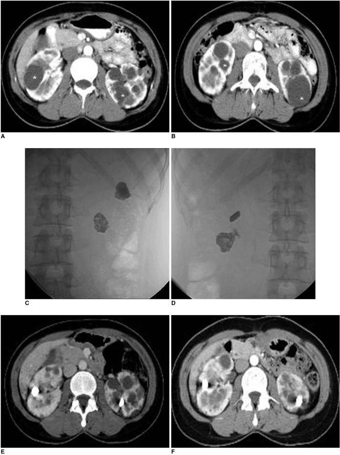
Fig. 1.
36-year-old woman with autosomal dominant polycystic kidney disease.
A, B. Axial contrast enhanced CT scans show multiple cysts in both kidneys. Cyst ablation was performed for two cysts in right kidney and for two cysts in left kidney (asterisk).
C, D. Antero-posterior radiographies after procedure show lobulated radiopacities that represent mixture of N-butyl cyanoacrylate and iodized oil in right (C) and left (D) kidneys.
E. Follow-up CT scans 36 months after procedure show collapsed cysts with mixture of N-butyl cyanoacrylate and iodized oil.
F. Follow-up CT scans at 60 months show no evidence of reaccumulation in ablated cysts.

