
Fig. 2. Delivery of siRNA by polymersomes.
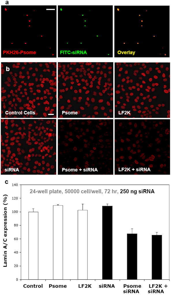
a) Fluorescent micrographs of fluorescent PKH26 labeling the membrane of nano-sized OLA polymersomes (Psome) and FITC-labeled siRNA for lamin A/C. The precise overlay of these two images indicates the encapsulation of siRNA in the aqueous lumen of OLA polymersomes. Scale bar = 10 μm. b) Representative images of A549 cells fixed and stained for lamin A/C at 72 hours display the knockdown of Lamin A/C in cultures incubated with LF2K-encapsulated siRNA and OLA-Psome encapsulated siRNA when compared to controls. Scale bar = 40 μm. c) Quantification of lamin A/C immunofluorescence in A549 cells indicates a 33-35% knockdown of lamin A/C by LF2K-encapsulated siRNA and OLA-Psome encapsulated siRNA.