
Figure 10.
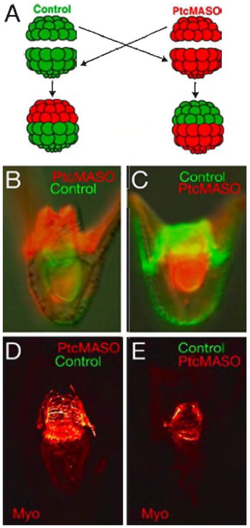
Reciprocal transplantation of animal and vegetal halves from control and Ptc MASO injected embryos (0.75 mM). (A) shows a diagram of the experiment. A control animal half (green) is placed on a red Ptc MASO-injected vegetal half, and reciprocally, a red Ptc-MASO-injected animal half is placed on a green control vegetal half. 48 hr embryos in which the Ptc MASO was in the animal half (B,D) and reciprocally, in which the Ptc MASO was in the vegetal half (C,E). The fluorescent lineage tracers are shown in (B,C), and (D,E) show the circumesophageal muscle of the embryo immediately above them as seen by confocal microscopy. When the Ptc MASO is in the vegetal half the muscle patterning is abnormal (E), while if the Ptc MASO is in the animal half the muscle pattern is normal (D).