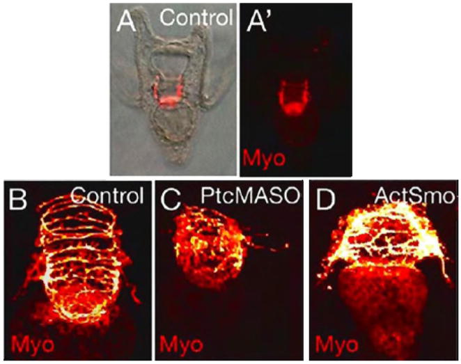
Figure 8.
Myosin antibody staining shows circumesophageal muscle organization in 48 hr control embryos (A,A′,B). In 48 hr embryos injected with Ptc MASO (0.75 mM) (C), and in 48 hr embryos expressing ActSmo (0.63 pg/pl)(D), the circumesophageal muscle is patterned abnormally compared to the control. Images are captured by DIC (A), fluorescence microscopy (A′), and by confocal microscopy (B–D).

