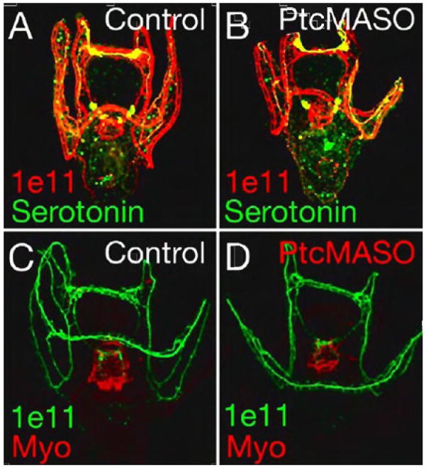
Figure 9.
Patterning of serotonergic and non-serotonergic neurons throughout the larvae shown by confocal projections of fluorescent nerve markers. Anti-serotonin (green) shows a normal pattern of serotonergic neurons in both control (A) and Ptc MASO injected embryos (B). Anti-Synaptotagmin B shows all neurons in red in control embryos (A) and in Ptc MASO injected embryos (B) and in green in (C, D). Myosin antibody staining in red (C,D) shows a normal pattern of circumesophageal muscle in the pharynx (C) and an abnormal pattern in the Ptc MASO injected embryos (D).

