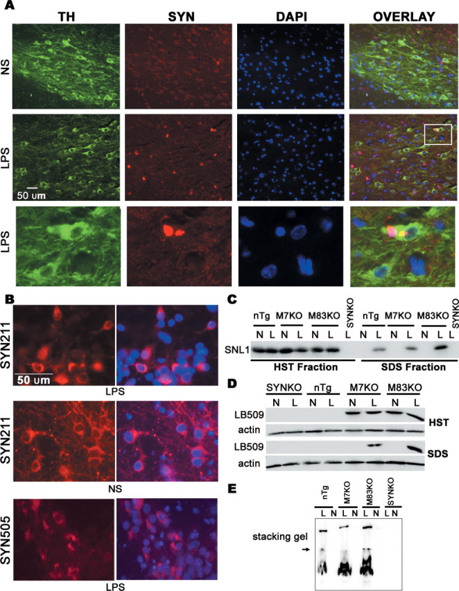
Figure 6.

Accumulation of insoluble and aggregated SYN in mouse midbrain after LPS injection. A, Mouse brain sections were double labeled with anti-TH (green) and SYN211 (specific to human SYN, red) antibodies. SYN211 staining revealed that some of TH-expressing cells in the LPS-injected SN contained SYN aggregates. The inset and its magnified photomicrograph (bottom) display SYN aggregates in TH-expressing neurons in the LPS-injected SN. B, In neuron–glia cultures, SYN appeared mainly in perinuclear locations and formed aggregation 7 d after the LPS treatment. SYN505, antibody raised to oxidized human SYN, positively stained these aggregates. C, D, Midbrain tissues were sequentially extracted and size fractionated by 12% SDS-PAGE gels, followed by Western blot analysis using antibody SNL1 or LB509. Insoluble SYN was detected only in extracts of LPS-injected midbrains, but not in NS-injected midbrains. E, HST-insoluble fraction (dissolved in RIPA buffer) was size fractionated on a nondenaturing 12% polyacrylamide gel and probed by Western blotting for SNL1. The aggregated SYN was seen in the stacking gel in LPS-injected midbrain extracts, but not in NS-injected midbrain extracts. The arrow indicates the resolving and stacking gel interface. N, NS; L, LPS; DAPI, 4′,6′-diamidino-2-phenylindole dihydrochloride.
