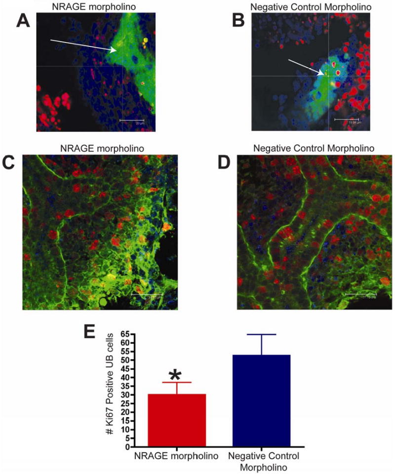
Figure 5. NRAGE expression mediates both apoptosis and proliferation in the developing Kidney.

(A-B) TUNEL analysis of E11.5 kidneys from the same Hoxb7-GFP embryo, one cultured with NRAGE morpholino (A) and the other kidney with negative control morpholino (B) for 72 hours (average projection of z-stack of all optical sections collected). Note (A-B): Green: ureteric bud (GFP), Red: TUNEL positive nuclei (TMR), Blue: all nuclei (TOPRO-3). (C-E) Proliferating cells were identified by whole mount Ki67 immunofluorescent staining of E11.5 kidneys, from the same ICR embryo, one cultured with antisense morpholino to NRAGE (C) and the other kidney with negative control morpholino (D) (optical section 2 μm). (E) There were significantly fewer Ki67 positive cells in the ureteric bud (*unpaired two-tailed t-test p=0.0024) in NRAGE morpholino treated explants in comparison to negative control treated explants. Note (C-D): Green: ureteric bud (Dolichos Bifluorous Aggultinin-Alexa488), Red: Ki67 positive nuclei (Ki67-Alexa546), Blue: all nuclei (TOPRO-3). Arrow: ureteric bud branches.
