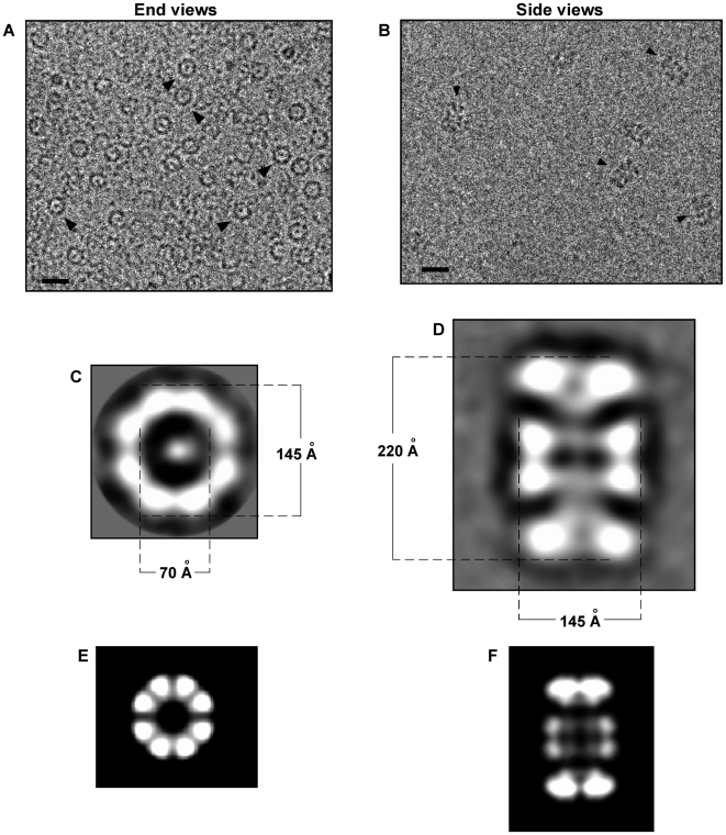
Figure 4. Cryo-electron microscopy of ssDNA-dependent oligomeric Rep68 rings.
Rep68-ssDNa complex was purified by size-exclusion chromatography in buffer A, and central part of the peak was concentrated and used for further cryo-electron microscopy analysis. (A) Representative image of the ssDNA-Rep68 oligomer; ring-shaped end view are shown by arrowheads. Bar corresponds to 20 nm. (C) A representative class average of end views is shown; internal and external dimensions of the ring are shown in Angstroms. (B) The ssDNA-Rep68 oligomer was purified by size-exclusion chromatography in buffer A; central part of the peak was concentrated and mixed with n-octyl β-D-glucopyranoside just before cryo-EM analysis. Arrowheads indicate side views of the Rep68 oligomer. Bar corresponds to 20 nm. (D) A representative class of side views is shown. Dimensions in Angstrom are shown for the length and width of the oligomer. (E and F) Two-dimensional projections of a double-octameric Rep68; end view (E), and side view (F).

