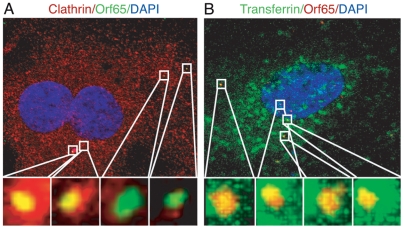
Figure 4. Colocalization of Orf65+ KSHV particles with markers of clathrin-mediated endocytosis during KSHV infection of endothelial cells.
(A) HUVEC were inoculated with KSHV, fixed at 1 hpi and stained for clathrin heavy chain (red) and Orf65+ viral particles (green). (B) HUVEC were infected with KSHV and simultaneously labeled with Alexafluor 488-transferrin (green). Cells were fixed at 1 hpi and stained for Orf65+ viral particles (red). Regions highlighted in the squares in the upper images are shown as enlarged 3D-projection images in the lower panels. Areas of colocalization of red and green signals are revealed as yellow. For corresponding 3D projection of colocalization of Orf65+ particles with clathrin, see Videos S5, S6, S7 and S8. For corresponding 3D projection of colocalization of Orf65+ particles with transferrin, see Videos S9, S10, S11 and S12.

