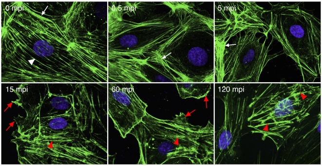
Figure 6. KSHV infection of endothelial cells induces actin dynamics and rearrangements of the actin cytoskeleton.
HUVEC were infected with KSHV and stained for actin cytoskeleton (green) and nuclei (blue) at different time points post-infection. Actin stress fibers (while arrow head) and cortical actin structures (white arrow) were visible before viral infection. KSHV infection disrupted actin stress fibers and induced more cortical actin structures at the early stage of infection (<15 mpi). However, cortical actin structures started to dissolve accompanying the reappearance of short and thick actin filaments resembling actin tails/spikes (red arrow head) after 15 mpi. Membrane ruffling, lamellipodia and filopodia were visible after 15 mpi (red arrow head).

