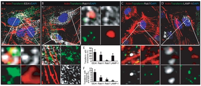
Figure 11. Association of Alexafluor 488-transferrin with actin filaments at different stages of endocytosis in endothelial cells.
(A–D) HUVEC were treated with Alexafluor 488-transferrin for 60 min, fixed and stained for actin filaments (red), markers of endosomes (white) and nuclei (blue). Alexafluor 488-transferrin shown in green was clearly associated with actin filaments and endosomal protein EEA1 (A), Rab11 (B), Rab7 (C) and LAMP-1 (D). The sections highlighted by the squares are enlarged in the lower or right panels. For corresponding 3D projection of colocalization of Alexafluor 488-transferrin with actin filaments and EEA1, Rab11, Rab7 and LAMP-1, see Videos S16, S17, S18, S19 and S20. (E–F) Quantification of colocalization of Alexafluor 488-transferrin with markers of endosomes (E), and with both actin filaments and markers of endosomes (F).

