
Figure 2.
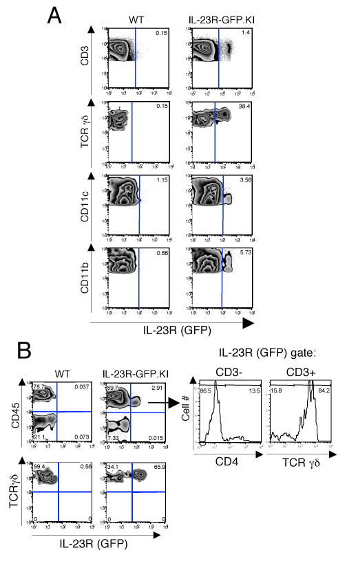
IL-23R expressing cells are enriched in the lamina propria (LP) of naive mice. Cells from lymph nodes, spleen and LP were prepared from naive WT and IL-23R-GFP.KI mice, stained and analyzed by flow cytometry. A, Lymph node cells were stained for CD3, TCRγδ, CD11c, and CD11b. B, IL-23R(GFP) expression was also analyzed in LP cells. LP cells were stained for CD45 and TCRγδ (B left panel). The expression of CD3, CD4, and TCRγδ was analyzed in the CD45+IL-23R/GFP+ compartment of LP cells (B right panel).