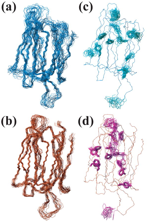
Fig. 2.

Three-dimensional NMR structure of At3g16450.1. (a) Superposition of the 20 energy-minimized conformers that represent the three-dimensional solution structure of the N-terminal domain. (b) Superposition of conformers representing the C-terminal domain. (c) Aromatic side chains (light green) and one backbone trace (blue) of the NMR structures for the N-terminal domain. (d) Aromatic side chains (magenta) and one backbone trace (red) of the NMR structure of the C-terminal domain.
