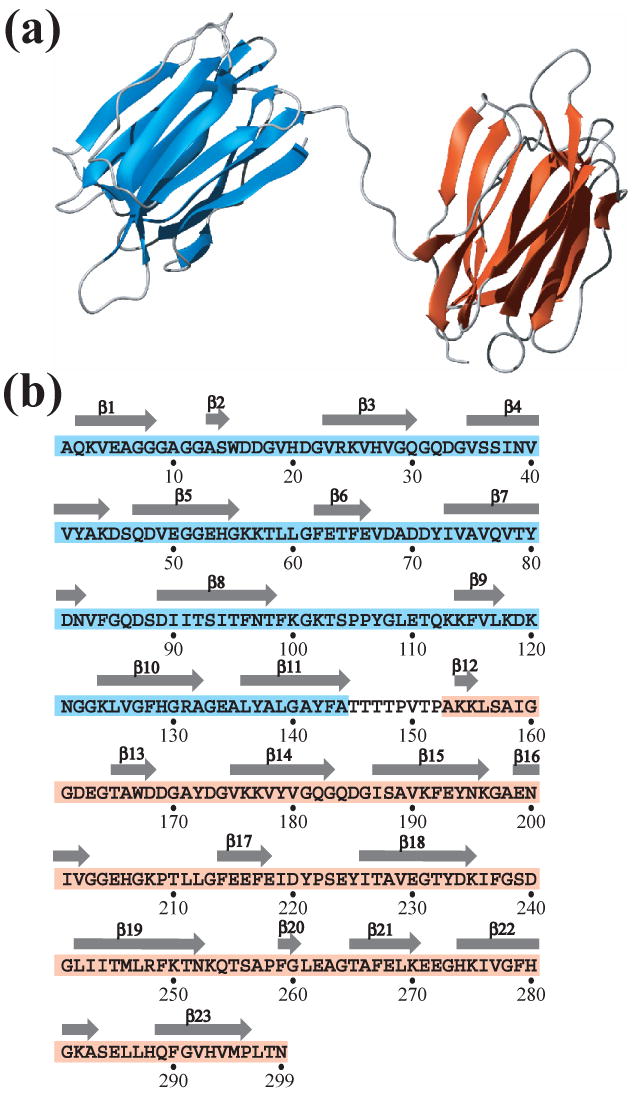
Fig. 3.

Secondary structure of At3g16450.1. (a) Ribbon representation of the NMR structure of At3g16450.1. These figures were prepared with MOLMOL (25). Due to the lack of NOEs, the relative orientation between the N- and C-terminal domains could not be defined. (b) Primary sequence of At3g16450.1. The sequences that correspond to the N-terminal (residues 1-144) and C-terminal (residues 153-299) structural domains are highlighted in cyan and pink, respectively, and β–strands are depicted as arrows above the sequence.
