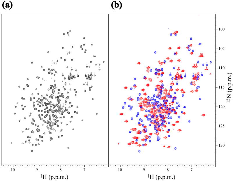
Fig. 4.
Comparison of the NMR spectra of full-length At3g16450.1 and its isolated N- and C-terminal halves. (a) 1H-15N HSQC spectrum of full-length (residues 1–299) SAIL-At3g16450.1. (b) Overlay of 1H-15N HSQC spectra of the N-terminal (residues 1–153, blue) and the C-terminal (residues 151–299, red) halves of [U-15N]-At3g16450.1. These spectra were acquired at 27.5 °C, pH 6.8 on a Bruker DRX600 NMR spectrometer. The pattern of the overlaid spectra is almost identical to that of full length construct, showing that the two domains of At3g16450.1 are largely independent.

