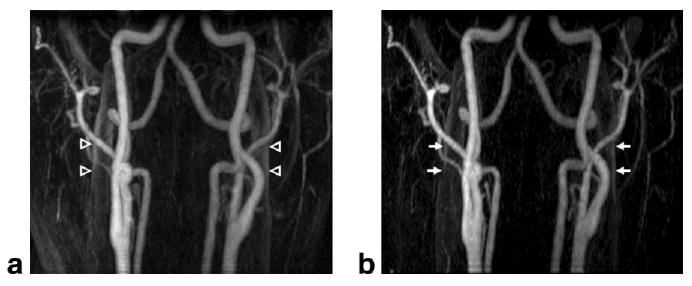
FIG. 4.

Results from volunteer study 7, in which the SENSE scan was acquired first. Mask-subtracted MIP images are shown for (a) non-SENSE and (b) SENSE acquisitions. Areas of venous enhancement are noted in the (a) non-SENSE image (open arrowheads), while corresponding locations in the (b) SENSE image (arrows) show significant venous suppression of approximately 20%, and improved conspicuity of the arteries.
