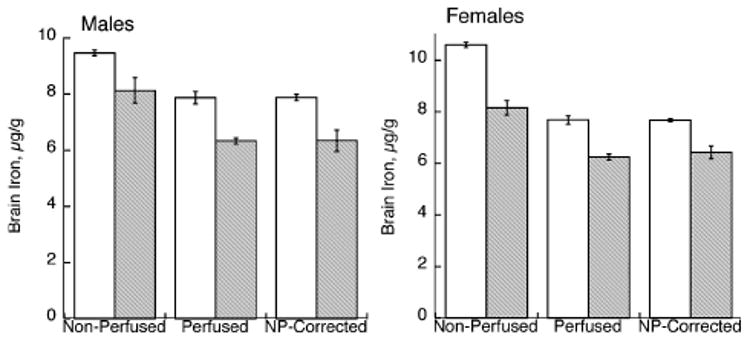
Fig. 2.

Effect of perinatal Cu deficiency on brain iron concentration in P24–P26 male and female rats. Iron was determined by flame atomic absorption spectroscopy in non-perfused and perfused rats. Each bar represents the mean ± SEM (n = 4). Open bars represent values for Cu-adequate rats and shaded bars for Cu-deficient rats. Perfusion lowered the brain iron concentration in both Cu-adequate and Cu-deficient rats of both genders, p < 0.05. Brain iron was determined in another set (n = 4) of non-perfused littermates but was corrected for blood Fe contamination after measurement of hemoglobin and estimation of blood iron. These values were not different from those measured in perfused littermates.
