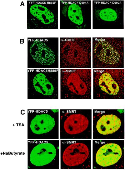
Figure 3.
Mutants of HDAC5 or 7 and the deacetylase inhibitor TSA or sodium butyrate disrupt the subcellular nuclear localization of HDAC5 and 7. (A) Direct fluorescence detection of mutant YFP-HDAC5 and YFP-HDAC7 in CV-1 cells shows the disruption of the characteristic subnuclear structures to a diffuse nuclear pattern. (B) Comparison of endogenous SMRT pattern when either wild-type YFP-HDAC5 or mutated YFP-HDAC5 is present CV-1 cells. The results are obtained with the use of direct and indirect immunofluorescence by using a SMRT antibody. Cells transfected with mutatedYFP-HDAC5H885F in contrast to wild-type-YFP-HDAC5 show a diffuse nuclear pattern that does not colocalize with endogenous SMRT in the nucleus. (C) Addition of the deacetylase inhibitors TSA and sodium butyrate disrupt the subcellular dot-like nuclear localization of HDAC5 and 7. Experiments were carried out by using direct fluorescence detection of wild-type YFP-HDAC5 in CV-1 cells after addition of TSA (100 nM; Upstate Biotechnology) or sodium butyrate (10 mM; Sigma). Images were viewed on an Olympus 1 × 70 inverted system microscope before deconvolation with deltavision2 software.

