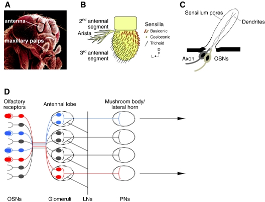
Fig. 1.
The olfactory system of Drosophila. (A) Drosophila melanogaster head. The white lines point to the olfactory appendages, the antenna and the maxillary palps. SEM image is courtesy of Jürgen Berger, Max Planck Institute for Developmental Biology, Tübingen, Germany. (B) Drawing of the three different classes of sensilla present on the 3rd antennal segment. The drawing is adapted with permission from Benton et al. (Benton et al., 2006). (C) Anatomy of a sensillum. Each sensillum is innervated by 2–4 olfactory sensory neurons (OSNs), which project an axon to the antennal lobe and dendrites into the sensillum lumen. The sensillum has pores that allow odorants to reach the neurons. (D) Organization of the Drosophila olfactory system. OSNs bind to odorants and send the information to glomeruli, innervated by local interneurons (LNs). The information then travels to higher centers in the brain, the mushroom body and lateral horn, through projection neurons (PNs).

