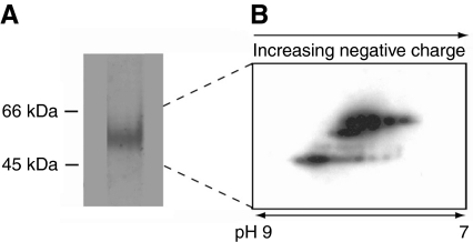
Fig. 10.
Thread matrix protein (TMP)-antibody cross-reactiivity at higher resolution. (A) Induced extracts separated on a one-dimensional sodium dodecyl sulfate polyacrylamide gel electrophoresis (SDS PAGE) gel and subsequently processed with anti-rTMP (only the relevant portion is shown). Proteins were typically not abundant enough for detection with Coomassie Blue stain. (B) Two-dimensional western blot (pH range ∼7–9) of the indicated proteins with anti-rTMP. Charge heterogeneity present in TMPs extracted from induced threads is evident as a laddering of spots. The 2-D blot has been enhanced so that the individual protein spots are clearly visible.

