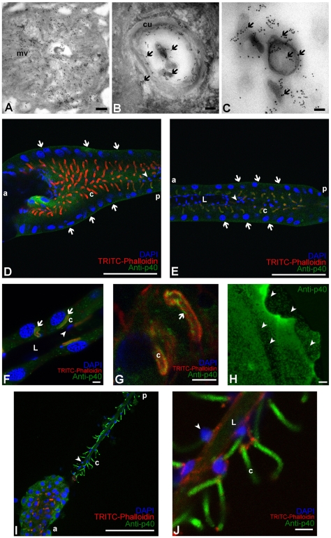
Fig. 4.
p40 localization in L. heterotoma venom gland. (A) Transmission electron micrograph with anti-p40 immunogold staining of the rough canal in cross section. p40 (black dots) is localized in the microvillar area (mv). Scale bar, 0.25 μm. Magnification: ×30,000, 80 KeV. (B) Transmission electron micrograph with anti-p40 immunogold staining of a smooth canal in cross section. p40 (arrows) is in the lumen of the canal, close to the walls and in the center, p40 is associated with larger electron-dense virus-like particle (VLP) precursors. cu, cuticle lining the canal. Scale bar, 90 nm. Magnification: ×85,000, 80 KeV. (C) Transmission electron micrograph of anti-p40-labelled section of the lumen of the venom gland. p40 (arrows) signal is predominantly around assembling VLPs. Scale bar, 0.05 μm. Magnification: ×40,000, 80 KeV. (D) Confocal image of the anterior part of the venom gland. p40 (green) is found peripherally in the secretory cell cytoplasm around the nuclei (arrows), but the p40 signal in and around the canals is not as strong (c, red). Smaller nuclei of cells of the intimal layer (arrowhead) are visible in the center of the gland. The p40 signal in the body of the gland is from the gland lumen, which is positioned below the focal plane of this image. a, anterior; p, posterior. Scale bar, 100 μm. Objective: EC Plan-Neofluar ×40/1.30, oil DIC optics. (E) Confocal image of the posterior part of the venom gland. The secretory cells, cells of the intimal layer and the venom gland lumen (L) are labeled as in D. The p40 signal (green), also present within the cytoplasm of the secretory cells (arrows), is concentrated around the canals. a, anterior; p, posterior. Scale bar, 100 μm. Objective: EC Plan-Neofluar 40x/1.30, oil DIC optics. (F,G) Confocal images of the posterior part of the venom glands. p40 (arrows) is found in the lumen of the canals (F) or colocalizing with the F-actin (c, red; G). Labeling as in E. Scale bar, 5 μm. Objective: Plan-Apochromat ×63/1.4, oil DIC optics. (H) Confocal images of the venom fluid stained with anti-p40 antibody (green). The p40 signal has a spot-like pattern (arrowheads). Scale bar, 5 μm. Objective: EC Plan-Neofluar ×40/1.30, oil DIC optics. (I,J) Confocal images of a venom gland dissected from a cytochalasin-treated L. heterotoma. Labeling is as in E. In the anterior part, secretory cells are shrunk (I) and actin staining around the canals (c) is weak. In the posterior portion (higher magnification in J) secretory cells are not visible, whereas the entire canals (c) persist, even though the rough portion (corresponding to the microvillar region) is not evident. a, anterior; p, posterior. Scale bar, 100 μm. Objective: EC Plan-Neofluar ×40/1.30, oil DIC optics. (J) Confocal image of a venom gland dissected from a cytochalasin-treated L. heterotoma. p40 (green)-filled canals (c) hang from the gland lumen (L), which is outlined by cells of the intimal layer (arrowhead). Labeling as in E. Scale bar, 5 μm. Objective: EC Plan-Neofluar 40x/1.30, oil DIC optics.

