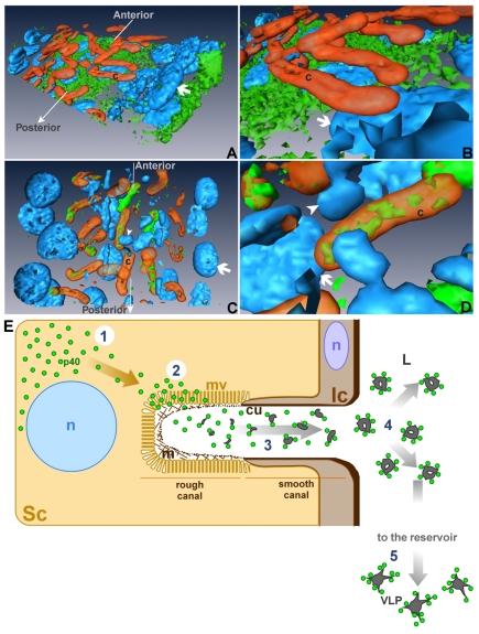
Fig. 5.
Model of synthesis and secretion of p40. (A,B) Three-dimensional reconstruction of a section of the anterior region of an L. heterotoma venom gland. p40 (green) is mainly localized to the outermost part of the secretory cells around nuclei (arrow). Actin canals (c) are positioned deeper in the cytoplasm where the concentration of p40 is lower. A close-up view (B) highlights the absence of p40 (green) within the canals (c) of this cell. (C,D) Three-dimensional reconstruction of a section of the posterior region of an L. heterotoma venom gland. p40 (green) is closely associated to the actin canals (c), either surrounding them or contained within their lumen. A close-up view (D) confirms that p40 (green) is found inside the lumen of some canals. The arrow points to a secretory cell nucleus; the arrowhead points to the nucleus of a cell of the intimal layer. (E) Organization of a secretory unit in the context of p40 synthesis, secretion and assembly of virus-like particles (VLPs). 1. p40 is synthesized in the perinuclear region of secretory cells (Sc). 2. p40 reaches the microvillar region (mv) of the canals where it is secreted into the canal lumen, passing through a putative extracellular mesh (m). 3. In the canal lumen, p40 associates with other components to assemble into VLP precursors. 4. VLP precursors containing p40 once released into the venom gland lumen (L), undergo additional structural changes. 5. Aggregates containing p40 travel along the gland lumen to reach the reservoir, where they mature into VLPs. Ic, cell of the intimal layer; n, nuclei; cu, cuticle.

