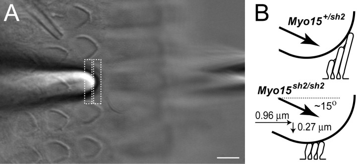
Figure 1.
Recordings of MET responses in cochlear hair cells. A, Cultured organ of Corti explant with a piezo-driven probe (left) and a patch pipette (right). A third pipette (top left) was used to deliver test solutions to the hair bundle. The image of the probe was projected to a pair of photodiodes (dashed rectangles) that monitor displacement stimuli. The specimen was visualized on an inverted microscope. Scale bar, 5 μm. B, In-scale schematic drawing of a 5 μm probe deflecting stereocilia of Myo15 +/sh2 (top) and Myo15 sh2/sh2 (bottom) IHCs. Stereocilia of Myo15 sh2/sh2 IHCs are deflected by the probe located at the top of the bundle.

