
Figure 2.
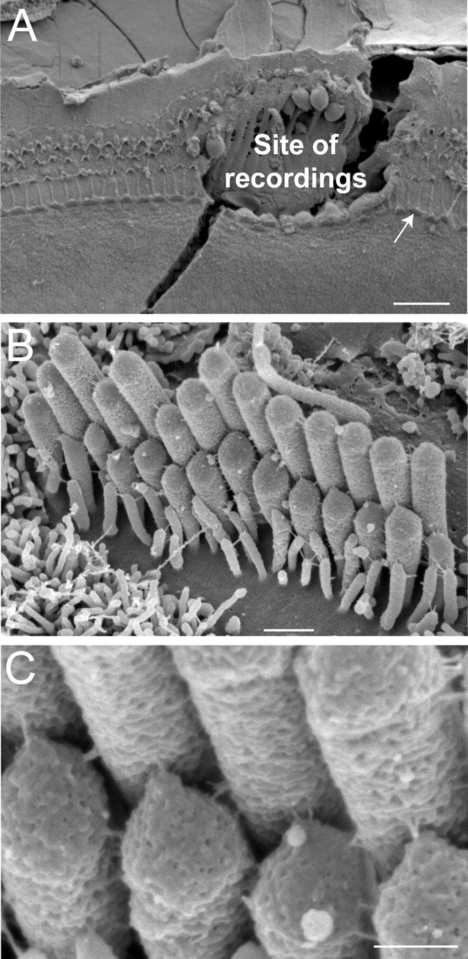
Postexperimental examination of specimens. A, The site of patch-clamp recordings is easy to identify even in the presence of the SEM preparation artifact (a fracture across the recording site). B, C, An IHC that is indicated by an arrow in A is shown at high (B) and very high (C) magnifications. Our oldest Myo15 +/sh2 preparation (P4 + 5 d in vitro) is shown to illustrate preservation of IHC morphology in vitro. Scale bars: A, 20 μm; B, 0.5 μm; C, 200 nm.