Figure 2.
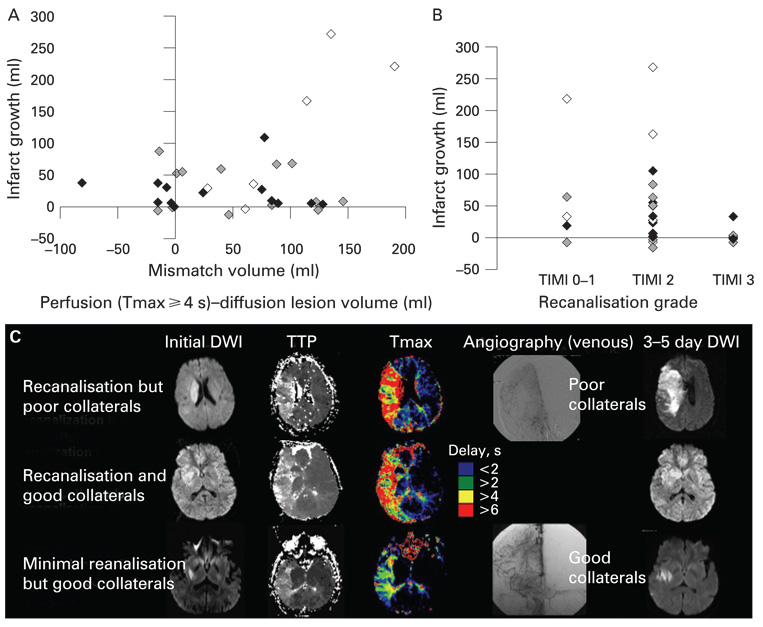
Impact of angiographic collateral grade on infarct growth. Displayed are scattered plots of (A) pretreatment diffusion–perfusion mismatch volume versus infarct growth and (B) the degree of recanalisation after treatment versus infarct growth. Filled diamonds indicate patients with good collaterals (grade 4), grey diamonds those with intermediate collaterals (grade 2–3) and open diamonds those with poor collaterals (grade 0–1). (C) Pretreatment diffusion and perfusion MRI findings and final diffusion weighted imaging (DWI) findings of patients with (i) recanalisation (Thrombolysis in Myocardial Infarction scale (TIMI) 2) but poor collaterals (grade 1), (ii) recanalisation (TIMI 3) but good collaterals (grade 4) and (iii) minimal recanalisation (TIMI 1) but good collaterals (grade 4). The initial DWI lesion volume, time to peak (TTP) delayed perfusion volume and mismatch area was similar among the patients. However, the patient with good collaterals showed minimal/no marked infarct growth, regardless of the occurrence of recanalisation, whereas infarct growth with clinical deterioration was observed in the patient with poor collaterals.

