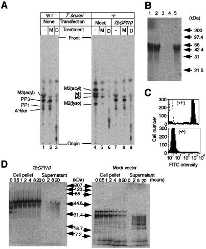
Figure 4.
Defective GPI biosynthesis, GPI anchoring, and surface expression of procyclins in TbGPI10 knockout procyclics. (A) GPI biosynthesis. Wild-type (WT) and doubly disrupted mutant (−/−), which were transformed with an empty vector (Mock) or TbGPI10 plasmid (TbGPI10), were used. The membranes were incubated with GDP-[3H]mannose to label GPI, and aliquots were subjected to TLC directly (−) or after digestion with α-mannosidase (M) or GPI-PLD (D). Identities of mannolipids are shown on the left of chromatograms. Designations of mannolipids from TbGPI10-disruptant are tentative. M1 and M2, intermediates containing one and two mannoses; M2(acyl) and M2(lyso), M2 species with acylation on inositol and with a lack of sn-2 fatty acid; M3(acyl), an intermediate bearing three mannoses with acylation on inositol; A′-like, an intermediate bearing three mannoses with ethanolamine phosphate on the third mannose; PP3, A′-like intermediate with acylation on inositol; PP1, complete GPI precursor (a lyso form of PP3). The spots that appeared after GPI-PLD-treatments (lanes 3, 6, and 9) are inositol-acylated GPI glycans. (B) Incorporation of myristic acid into procyclins. Lane 1, wild-type; lane 2, single TbGPI10-disruptant; lane 3, double TbGPI10-disruptant; lane 4, double TbGPI10-disruptant bearing an empty plasmid; lane 5, double TbGPI10-disruptant bearing TbGPI10 plasmid. Size markers are on the right. (C) Surface expression of EP procyclins. Single and double TbGPI10-disruptant clones were stained with anti-EP procyclins (shaded lines) or control (dotted line) monoclonal antibodies and analyzed in a FACScan. (D) Pulse-chase analysis of EP procyclins. Double TbGPI10-disrupted mutant bearing TbGPI10 (Left) or empty (Right) plasmid was pulse-labeled with [14C]proline for 30 min and chased for indicated time periods. At each time point, aliquots of samples were separated into supernatants and cell pellets, solubilized by detergent, and immunoprecipitated with anti-EP procyclins antibody. Immunoprecipitates were analyzed by SDS/PAGE and autoradiography.

