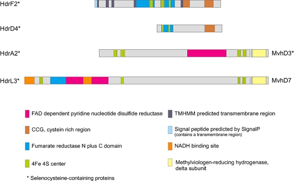
Fig. 5.

Pfam (domain scans analysis) and domain alignments of four types of heterodisulfide reductase subunits (Hdr) of Db. autotrophicum HRM2. Relevant Pfam domains are given in colour codes. All Hdr proteins depicted contain selenocysteine; the HdrA and the HdrL type of the proteins are colocated with methylviologen non-reducing hydrogenase subunit D (mvhD). The deduced HdrF protein contains a domain with several transmembrane regions indicating a possible membrane integration of the protein. In contrast, HdrD, HdrA and HdrL have domain structures typical for soluble proteins.
