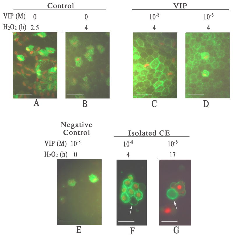
Fig. 2.
Fluorescence microscopy of flat-mounted corneoscleral explants showing VIP pretreatment promoted apoptosis (annexin V-binding) at the expense of necrosis (PI-staining) among CE cells injured by severe oxidative stress. (A & B) Necrotic CE cells in a control cornel cup 2.5 h (A) and 4 h (B) after H2O2 treatment. With compromised cell membrane, necrotic CE cells showed both PI-staining nuclei and annexin V-binding cell membrane. (C & D) Apoptotic CE cells appeared in VIP-pretreated corneal cups following 4 h of H2O2 treatment. 10−8 (C) and 10−6 (D) M VIP-pretreated corneal cups showed numerous annexin V-binding only (no PI-staining) apoptotic CE cells. (E) Negative control for apoptosis. No annexin V-binding only apoptotic CE cell was observed in 10−8 M VIP-pretreated corneal cup that was not subjected to subsequent H2O2 treatment. (F &G) Annexin V-binding to apoptotic CE cell membranes free of cell-cell junctions (arrows). CE cells were isolated from corneal cups that have been pretreated with 10−8 M (F) and 10−6 M (G) VIP followed by 4 h (F) and 17 h (G) of H2O2treatment, respectively. White bar length=15 μm.

