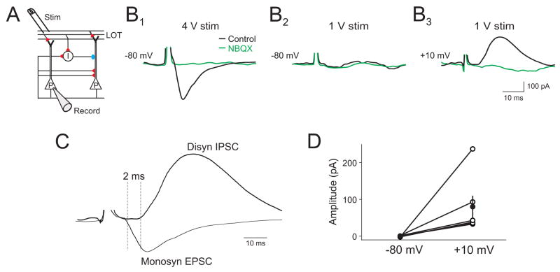
Figure 5. Minimal stimulation of the LOT in vivo preferentially recruits disynaptic inhibition.
(A) Schematic of recording setup. (B1) Under control conditions, direct LOT stimulation evokes a monosynaptic EPSC (Vm=−80 mV) at high stimulation intensity (4 V) in a L2/3 cell. (B2) Lowering stimulation intensity (1 V) fails to evoke an EPSC, while depolarization to +10 mV reveals an IPSC (B3). Subsequent application of NBQX (500 μM) to the cortical surface abolishes the monosynaptic EPSC and disynaptic IPSC (B1–3, green traces). (C) Overlay of monosynaptic EPSC and disynaptic IPSC. (D) Summary data of recruitment of disynaptic IPSCs (+10 mV) at stimulus intensities that failed to evoke EPSCs (−80 mV, n=5 cells).

