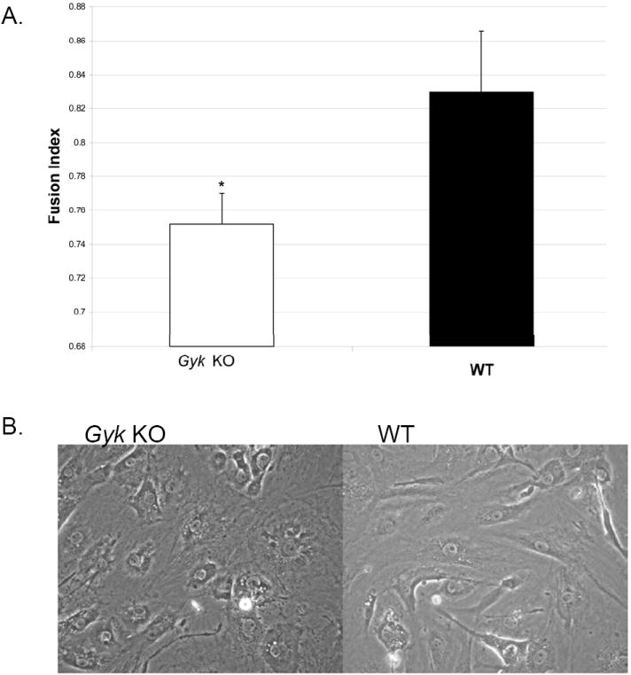
Figure 5.

Myotube fusion index of Gyk KO and WT primary muscle cells: A. fusion index measured as the ratio of the number of nuclei in myotubes (3 or more nuclei) to the total number of nuclei counted of day 6 after the addition of fusion media as described in Materials and Methods. *p value <0.05, student t test. Black bar represents WT muscle cells and white bar represents Gyk KO muscle cells. B. Photograph of WT and Gyk KO muscle cells on day 5 after supplemented with fusion media.
