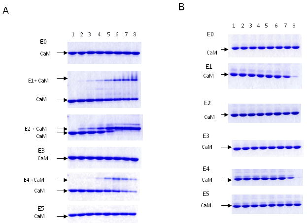
Fig.3. The interaction of peptides based on eNOS sequence with CaM determined by gel mobility shift.

CaM (200 pmole) was incubated with each peptide (E0, E1, E2, E3, E4, E5) by increasing CaM:peptide molar ratio for 1 h. The samples were electrophoresed on 18% nondenaturing gels in the presence of 100 μM Ca2+ (Panel A), or 1 mM EGTA (Panel B). The free CaM and CaM/peptide complex were visualized by Coomassie-blue R250 and indicated. The lane 1 in each gel contains CaM alone. CaM:peptide ratios used were: lane 2 (1:0.15), lane 3 (1:0.3), lane 4 (1:0.5), lane 5 (1:1), lane 6 (1:2), lane 7 (1: 5) and lane 8 (1:10).
