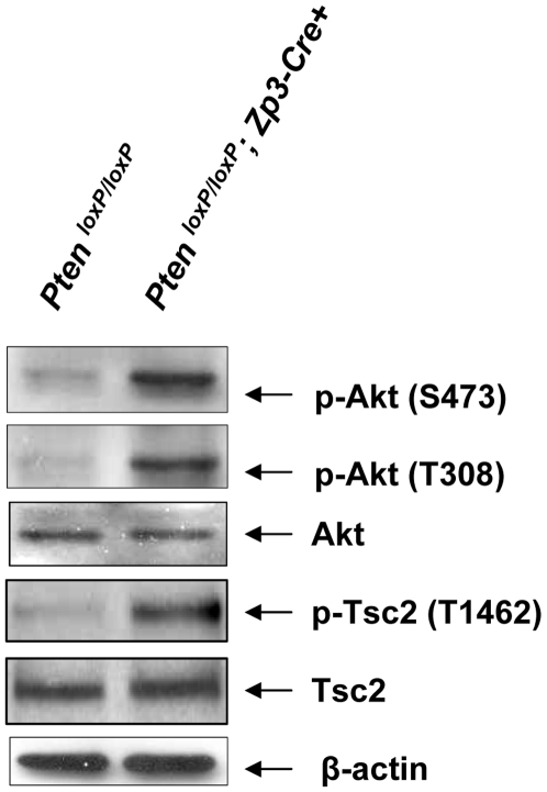
Figure 3. Enhanced Akt signaling in oocytes of PtenloxP/loxP; Zp3-Cre+ mice.
Oocytes were prepared from ovaries of 3–4 week old mice that were treated with PMSG, as described in Materials and Methods . Signaling studies in PtenloxP/loxP; Zp3-Cre+ oocytes showed elevated levels of p-Akt (Ser473), p-Akt (Thr308), and p-Tsc2 (Thr1462) as compared to PtenloxP/loxP oocytes. Levels of total Akt, Tsc2, and β-actin were used as internal controls. 100–150 oocytes were used for each lane. All experiments were repeated at least three times and representative results are shown.

