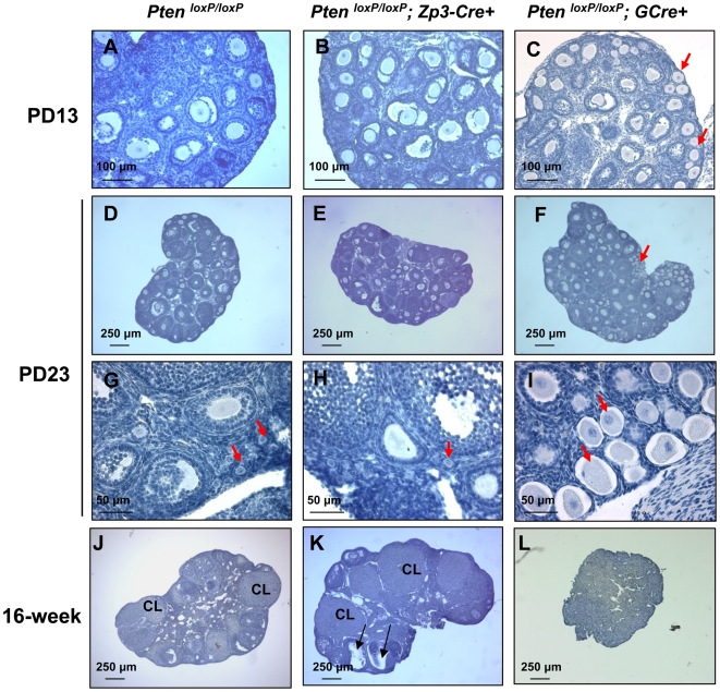
Figure 4. Normal follicular development in PtenloxP/loxP; Zp3-Cre+ mice.
Morphological analysis of ovaries from 13- and 23-day-old, and 16-week-old PtenloxP/loxP; Zp3-Cre+ mice, PtenloxP/loxP; GCre+ mice, and control PtenloxP/loxP mice. Ovaries were embedded in paraffin and sections of 8-µm thickness were prepared and stained with hematoxylin. Note the overactivation of primordial follicles in PtenloxP/loxP; GCre+ ovaries (C, F, and I, arrows) and the normal follicular development and CL in PtenloxP/loxP; Zp3-Cre+ ovaries (B, E, H and K), which is comparable to the control PtenloxP/loxP ovaries (A, D, G, and J). CL, corpora lutea.

