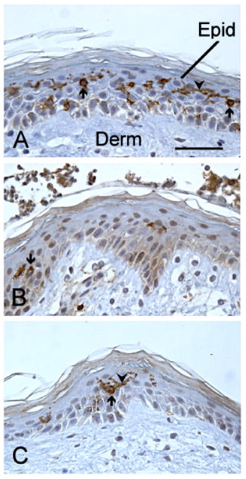
Figure 3.
Cross-sections of human skin grafted to the chick chorioallantoic membrane and stained with anti-CD1a to reveal LC. The sections are counterstained with hematoxylin to reveal the general structure of the skin. Panel A shows a control skin fragment treated overnight with solvent (PBS). A few of the many brown-stained LC are designated by arrows, and some of their processes are indicated by arrowheads. In B, a fragment treated with DNFB is shown. One LC, and a few stained processes can be seen. C shows a fragment treated overnight with nLT, with a single, large LC, with long process. Epid- epidermis, Derm- dermis, calibration bar = 50mm.

