
Fig. 2.
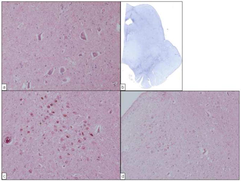
Neuropathologic findings from 2 subjects (cases 3 and 7) with PARK8 parkinsonism in the current study. A, View of the substantia nigra in case 3. Neuronal cell loss and gliosis were mild compared with that seen in sporadic Parkinson disease. (Hematoxylin-eosin; original magnification, ×200.) B, Scanning view of the midbrain in case 3. Marked gliosis was seen in the area of the substantia nigra, especially the pars reticulata. (Holzer stain.) C, View of the locus ceruleus in case 3. Neuronal cells are well preserved. (Hematoxylin-eosin; original magnification, ×100.) D, View of the locus ceruleus in case 7. Neuronal cells are well preserved. (Hematoxylin-eosin; original magnification, ×100.)