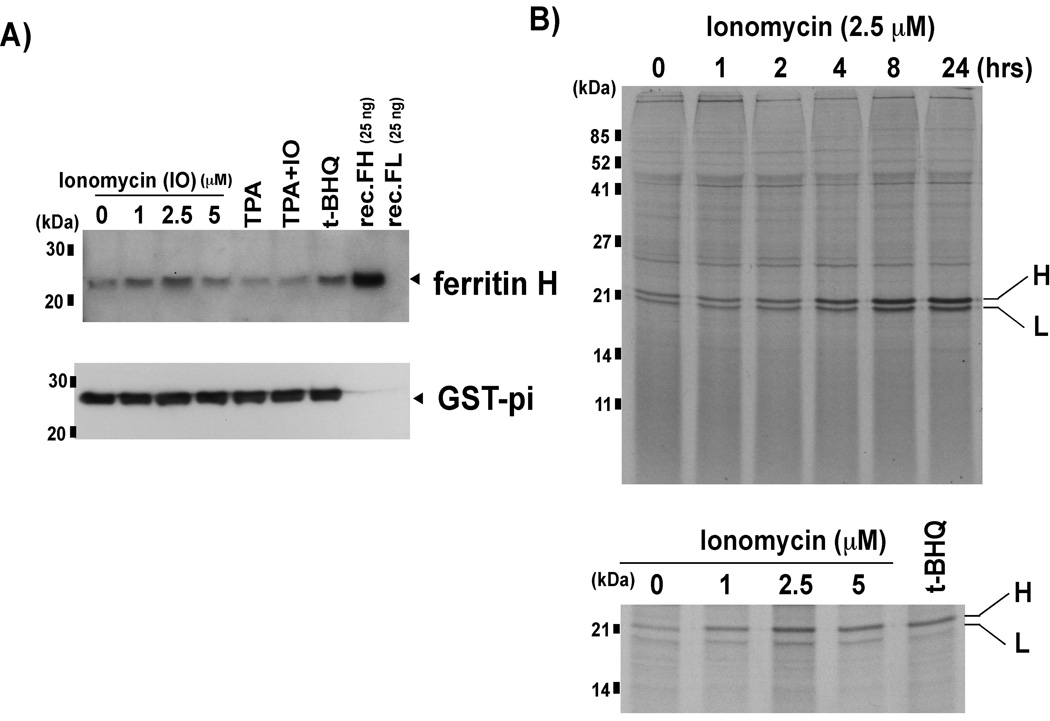
Fig. 1. Ferritin protein expression is increased by calcium ionophore treatment.
A) Jurkat cells were treated with 0, 1, 2.5 and 5 uM ionomycin (IO), 50 ng/ml TPA, 50 ng/ml TPA+ 2.5 uM IO, or 10 uM t-BHQ (as a positive control for ferritin H induction, [18]) for 24 h, and whole cell extracts were subjected to Western blotting using anti-Ferritin H antibody. Recombinant ferritin H and L (recFH, recFL) were included to assess the specificity of the antibody. Ferritin H band is indicated with an arrowhead. The membranes were re-probed with anti-GST-pi antibody. B) In vivo 35S methionine/cysteine labeling was performed following incubation with 2.5 uM ionomycin for 0–24 h (top), or 24 h incubation with 0–5 uM ionomycin (bottom). Lysates equaling 1×10⁷ TCA insoluble counts were subjected to immunoprecipitation with anti-ferritin antibody. Ferritin H and L bands are indicated (H, L).

