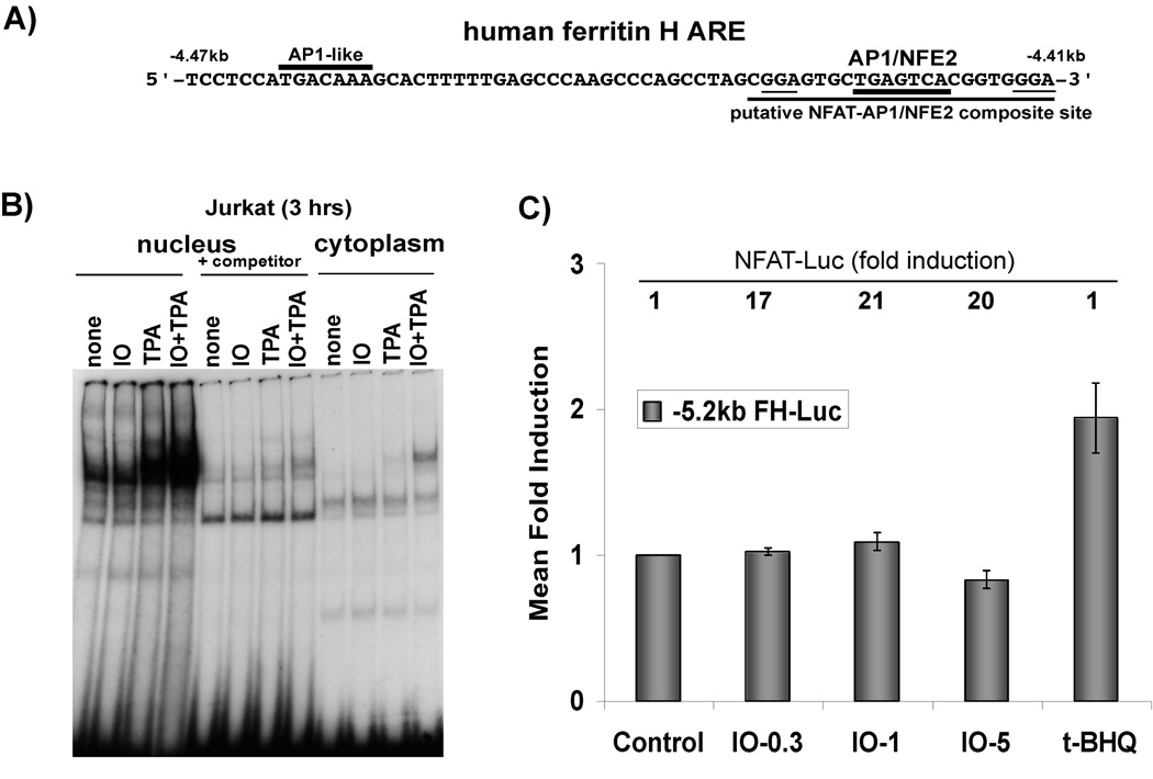
Fig. 4. NFAT is not involved in ferritin H transcriptional activation by calcium ionophore.
A) Schematic of the human ferritin H ARE sequences, including critical AP1-like, AP1/NFE2 sites, and putative NFAT binding sites (underlined). B) Nuclear and cytosplasmic extracts were prepared from cells treated with 2.5 uM ionomycin (IO), 50 ng/ml TPA, or 2.5 uM IO + 50ng/ml TPA for 3 h. 30 ug of each fraction was utilized for gel retardation assay with a γ32P-ATP labeled extended AP-1/NFE2 probe containing the putative NFAT site (underlined in A). +Competitor indicates the addition of the unlabeled NFAT oligonucleotide in 50-fold excess to the binding reaction. C) Jurkat cells were transiently transfected with −5.2 kb human FH-Luc and were allowed 24 hr recovery period. Then they were treated with DMSO control or 0.3, 1, or 5 uM ionomycin (IO), or t-BHQ (10 uM). Cell lysates were prepared and luciferase activity was assessed via luminometry with dual luciferase reagents (Promega). Luciferase activity in DMSO-treated cells was set to 1. S.E.M are shown, n=6 independent experiments with duplicate samples. The relative activity of NFAT-Luc (mean fold induction) for each treatment is indicated above each respective bar at the top of the graph.

