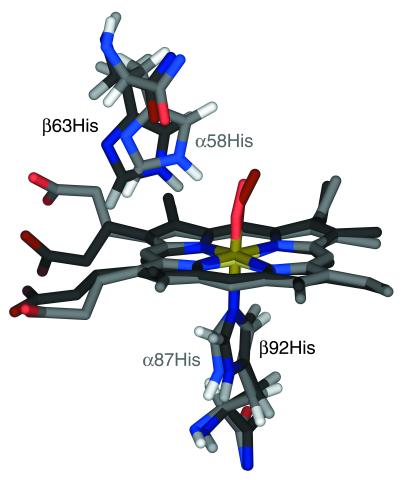
Figure 2.
Proximal and distal histidyls of the HbO2 A x-ray crystal structure (PDB entry 1HHO; ref. 8), with the hemes of the α- and β-subunits superimposed. The coordinates of hydrogen atoms were calculated from those of heavy atoms, using standard bond lengths and angles. The α- and β-subunits are shown in lighter and darker colors, respectively. This structure suggests that the distal histidine in the α-subunit is better disposed to form a H-bond with the O2 ligand than is its counterpart in the β-subunit.

