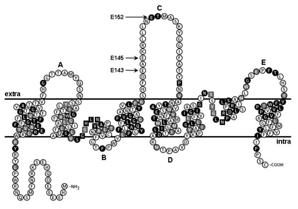
Figure 1.

Predicted topology of LmAQP1. Amino acids labeled as white letters in black circles are conserved between LmAQP1 and PfAQP; residues marked as white letters in gray circles are similar to PfAQP; black and gray squares indicate conserved and similar residues, respectively, which directly interact with glycerol in PfAQP. This topology was plotted using the TeXtopo package (Beitz, 2000).
