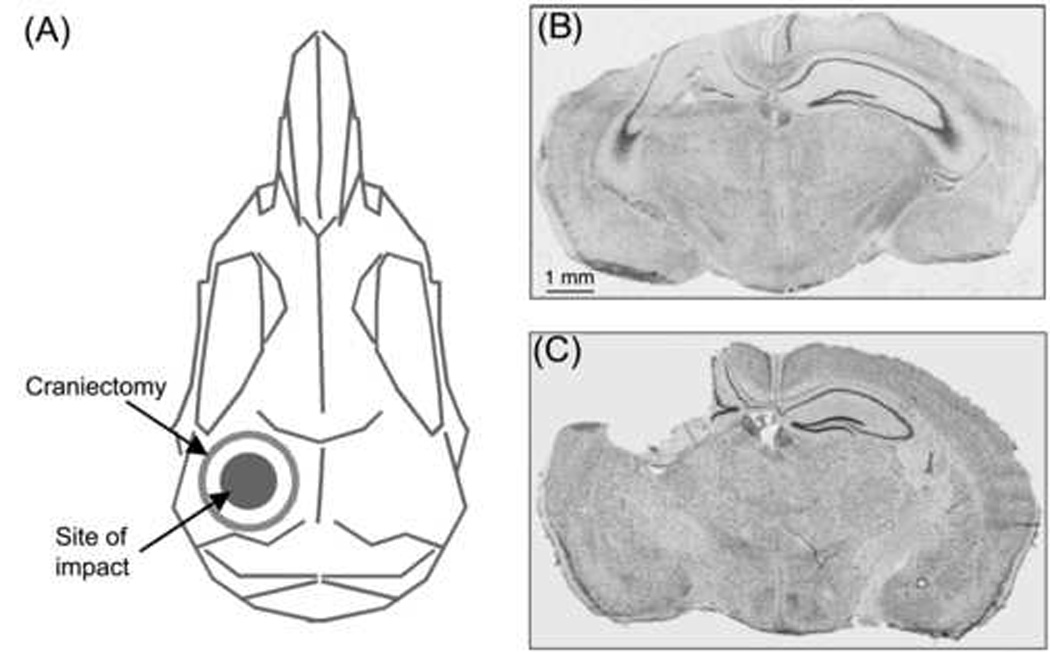
Figure 1.

(A) Schematic dorsal view of a mouse skull indicates site of craniectomy and impact. (B) Composite digital micrograph from a 5-month-old wild-type mouse (6 days after injury). In addition to direct cortical neuron loss at the injury site (on the left side of this section), there is ipsilateral loss of dentate granule cells and hippocampal pyramidal neurons. (C) Composite digital micrograph from a 20-month-old 4/4 apoE mouse (14 months after injury). The injured cortex and dorsal hippocampus had been cleared, presumably by macrophages, resulting in a large cavity at the injury site. Composite digital micrographs (B,C) were made from coronal sections stained for Nissl substance (thionin).
