
Figure 2.
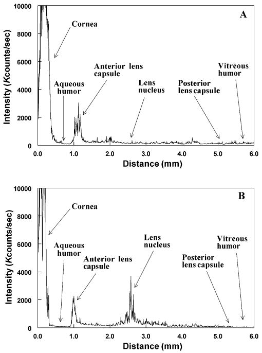
Typical static light scattering (SLS) analyses in vivo of eyes of control and UVA-treated guinea pigs. Each analysis was conducted along the optical axis of the lens, measuring 4.5–5.0 mm in diameter. Kcounts/s: 1000 photon counts per second of collected light intensity. (A) Control, 23-month-old untreated animal; (B) animal treated over a period of 5 months with UVA light. Note the increase in SLS intensity in the UVA-exposed lens, mainly in the central region, compared with that of an age-matched control animal.