
Figure 3.
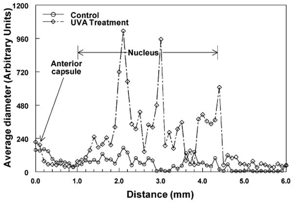
Representative dynamic light scattering (DLS) analysis profiles in vivo across lenses of control and UVA-treated guinea pigs. Fifty measurements of average protein diameter were made every 0.1 mm across the optical axis of each lens. The instrument made 10–15 measurements of protein diameter at each location and provided an average value. DLS data are not expressed as absolute sizes of lens proteins but as arbitrary units of diameter (see Materials and Methods for an explanation). (○) 23-month-old control animal; (⋄) 23-month-old experimental animal after 5 months of UVA light exposure.