
Figure 1.
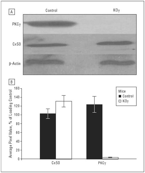
Western blots of whole retinal tissue. Whole retina from 6-week-old control or protein kinase Cγ– (PKCγ-) knockout (KOγ) mice were loaded in sample buffer at 40-μg protein per lane. A, Immunoblots using mouse anti-PKCγ antisera at 1:1000, mouse anti–connexin 50 (Cx50) at 1:500, and mouse anti–β-actin at 1:20 000. β-Actin is used as an internal loading control. B, The average pixel values of Cx50 and PKCγ in control and KOγ mice. Error bars indicate standard deviation.