
Figure 5.
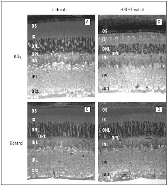
Structure of mouse retinas (aged 14 weeks) before and after hyperbaric oxygen (HBO) treatment. Sections of fixed retina, about 250 μm away from the optic nerve, are shown. GCL indicates ganglion cell layer; INL, inner nuclear layer; IPL, inner plexiform layer; IS, inner segments; KOγ, protein kinase Cγ knockout; ONL, outer nuclear layer, OPL, outer plexiform layer; OS, outer segments.