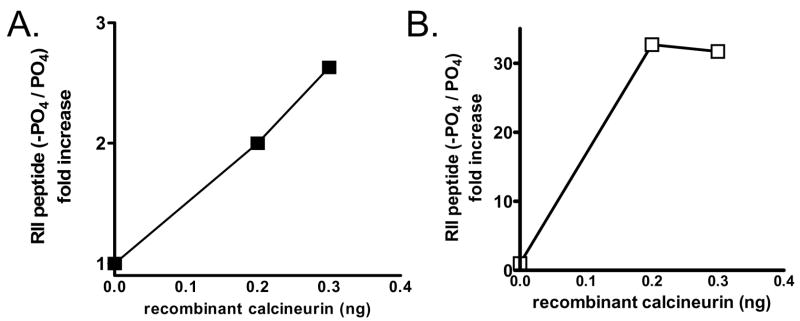
Figure 2. Validation of fluorescein-labeled RII peptide by mass spectrometry.
A) Reactions were carried out with 0, 0.2, or 0.3 ng of recombinant calcineurin per reaction and then the relative amount of dephosphorylated to phosphorylated peptide was determined by mass spectrometry. Data shown is the ratio of the area under the curve for dephosphorylated RII and phosphorylated RII with each condition. B) Reactions were carried identically as in (A) and then incubated with TiO4 matrix in a 96-well plate. After binding, the samples were removed and the relative amount of dephosphorylated to phosphorylated peptide was determined by mass spectrometry. Data shown is the ratio of the area under the curve for dephosphorylated RII and phosphorylated RII with each condition.

