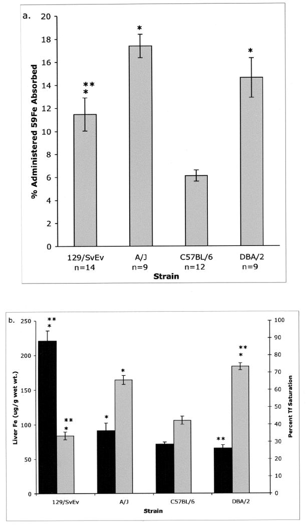
Figure 1. Iron measures in different strains of mice.

a. Mice were fasted and given a measured dose of 59Fe by gavage. Twenty-four h later, mice were sacrificed, dissected and the percent of the administered radiolabel remaining with the carcass minus the head and GI tract was measured. b. The hepatic iron content (black bars) and the percent Tf saturation (grey bars) were determined for the four strains. N values are the same for animals in Figures a and b. Error bars represent SEM.
* P< 0.05 vs. C57BL/6. ** P< 0.05 vs. A/J.
